New Nova 32-Channel Coil
Summary
CNI’s original Nova 32-channel coil went out of service earlier this year. After multiple discussions with Nova, it appears that the original coil cannot be fixed. We are fortunate that our friends at GE generously lent us a Nova coil that we have been using since the end of January.
We purchased and have received a new Nova 32-channel coil (the same model as currently in use). We conducted a number of tests validating that its performance matches our borrowed Nova coil. The results described here show a slight performance improvement (see below) for CNI’s new Nova coil compared to the one we borrowed. We have put this coil into operation effective as of today. GE’s Nova coil is being returned to them with many thanks.
SNR Comparison Between the two Nova Coils
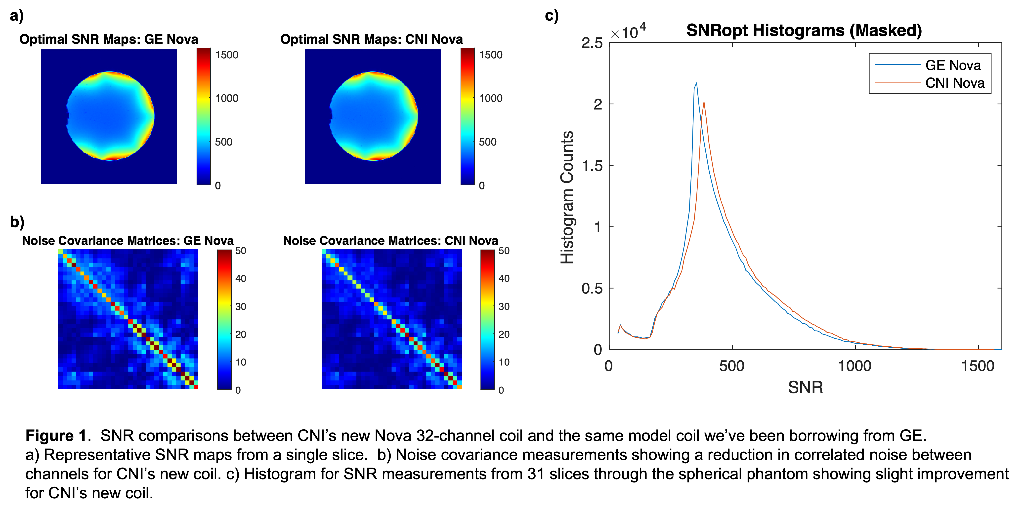
To compare the signal quality on the two coils, we acquired a multislice 2DFT sequence using a phantom. We then repeated the acquisitions with the RF flip angle set to 0 in order to acquire pure noise images. The SNR maps for an optimal coil combining (Roemer PB, et al. The NMR phased array. Magn Reson Med. 1990 Nov;16(2):192-225.) are shown for representative slices are c0mpared in Fig. 1a. The sensitivity shading and SNR maps are comparable between coils across all slices covering the volume.
The noise covariance arrays for the 32-channel data (Fig. 1b) with the new CNI Nova coil are somewhat more concentrated on the diagonal – a good thing meaning better coil independence. The signal-to-noise ratio (SNR) histograms from the voxels inside the phantom are also comparable (Fig 1c). The CNI Nova coil is slightly higher (histogram shifted to the right), but by an amount that is irrelevant compared to the impact of the physiological noise that will be present when scanning a brain.
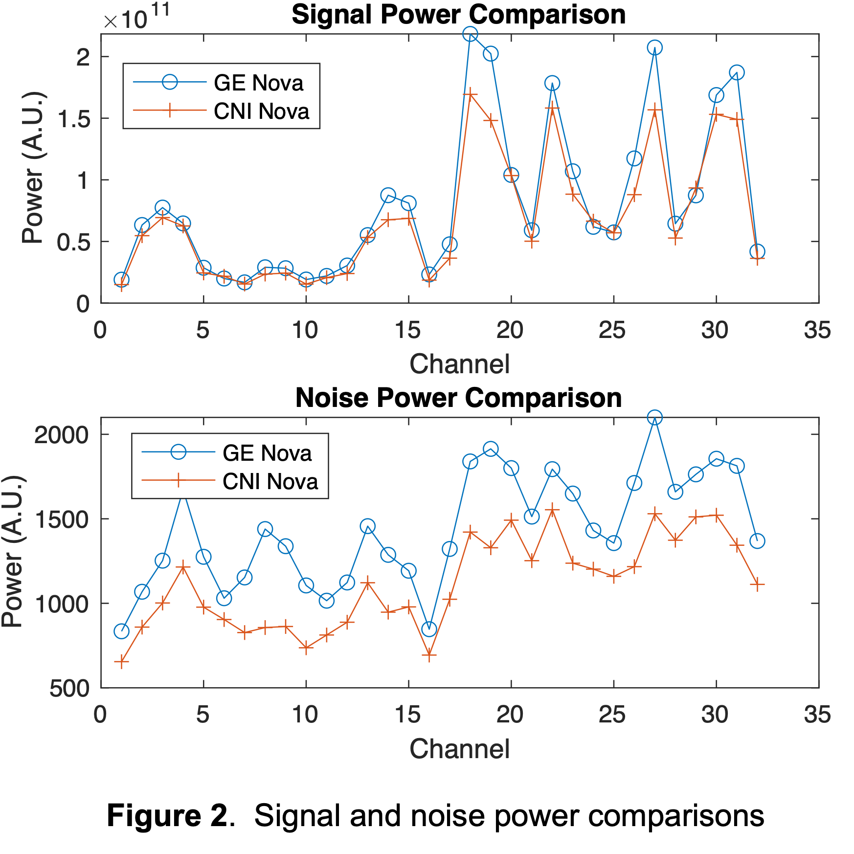
We also compared the integrated signal and noise power across the whole volume for each of the coil channels (Figure 2). The signal power follows the same trend across coils (individual elements are in the same positions and so we expect similar distribution of maxima and minima). The noise power values can also be impacted by local loading of the array by the phantom which leads to the similar higher distribution on the channels 17-32 which correspond to the lower (posterior) half of the array. We do not see any individual coil signal drops in the new CNI Nova coil, and that’s a good thing.
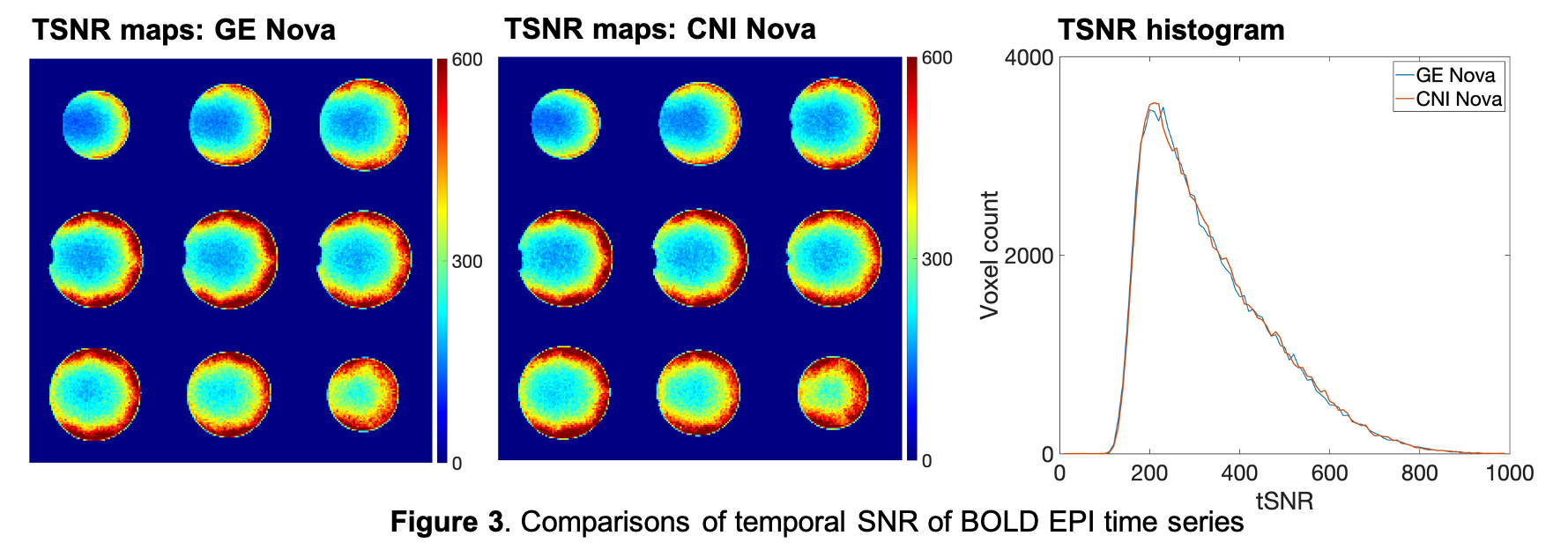
Finally, the temporal SNR of an axial BOLD EPI time series are very similar (Figure 3). The images on the left are tSNR maps and the graph on the right is the tSNR histogram from voxels in all 42 slices, covering the whole spherical phantom. There are no meaningful differences – especially compared to the additional physiological noise present in any real brain scan.
New Nova Coil Base
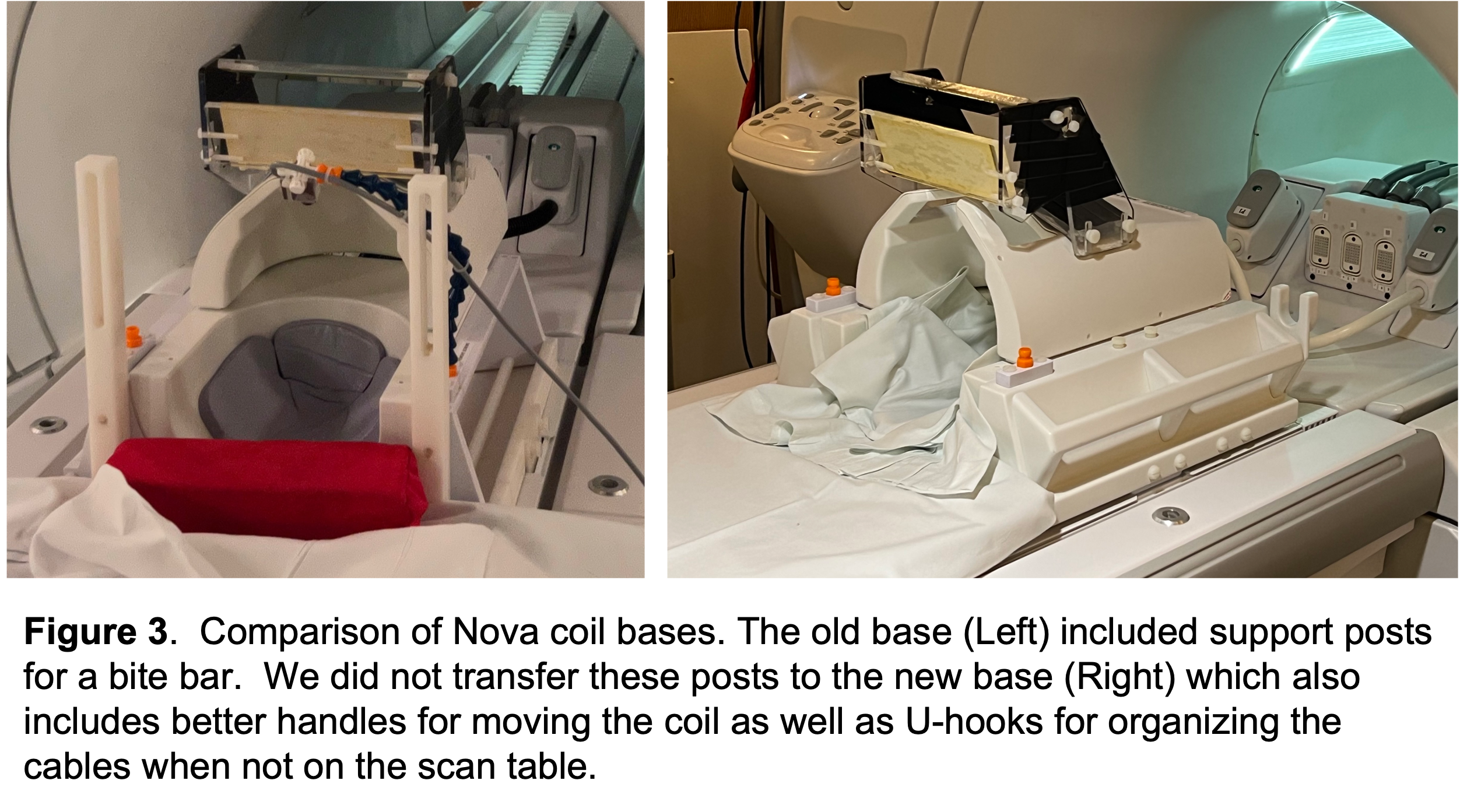
We’ve chosen to use the new base that came with our coil. The new base has better handles for transporting the coil, as well as cable management hooks. We’ve taken this opportunity to also abandon the bite bar posts and have not transferred them from the old base. The double mirror and Loc-line connectors for the webcam / IR illuminator / etc. were transferred.
Summary
We installed the newly purchased Nova coil. Its performance is very similar to the borrowed Nova coil we have been using. Thank you to GE! And to everyone else – please carry on as you were. We at the CNI are feeling grateful to our friends and glad to have this episode in our rear view mirror.
The CNI Team